In 2015 we provided outcomes showing that a deep knowing system (DLS) can be trained to examine external eye pictures and forecast an individual’s diabetic retinal illness status and raised glycated hemoglobin (or HbA1c, a biomarker that shows the three-month typical level of blood sugar). It was formerly unidentified that external eye pictures included signals for these conditions. This amazing finding recommended the possible to lower the requirement for customized devices because such pictures can be recorded utilizing smart devices and other customer gadgets. Motivated by these findings, we set out to find what other biomarkers can be discovered in this imaging method.
In “ A deep knowing design for unique systemic biomarkers in pictures of the external eye: a retrospective research study“, released in Lancet Digital Health, we reveal that a variety of systemic biomarkers covering numerous organ systems (e.g., kidney, blood, liver) can be anticipated from external eye pictures with a precision exceeding that of a standard logistic regression design that utilizes just clinicodemographic variables, such as age and years with diabetes. The contrast with a clinicodemographic standard works due to the fact that threat for some illness might likewise be examined utilizing an easy survey, and we look for to comprehend if the design translating images is doing much better. This work remains in the early phases, however it has the possible to increase access to illness detection and tracking through brand-new non-invasive care paths.
 |
| A design producing forecasts for an external eye image. |
Design advancement and examination
To establish our design, we dealt with partners at EyePACS and the Los Angeles County Department of Health Solutions to develop a retrospective de-identified dataset of external eye pictures and measurements in the type of lab tests and crucial indications (e.g., high blood pressure). We filtered down to 31 laboratory tests and vitals that were more typically offered in this dataset and after that trained a multi-task DLS with a category “head” for each laboratory and crucial to forecast irregularities in these measurements.
Significantly, examining the efficiency of lots of irregularities in parallel can be troublesome due to the fact that of a greater opportunity of discovering a spurious and incorrect outcome (i.e., due to the numerous contrasts issue). To reduce this, we initially assessed the design on a part of our advancement dataset. Then, we narrowed the note down to the 9 most appealing forecast jobs and assessed the design on our test datasets while fixing for numerous contrasts Particularly, these 9 jobs, their associated anatomy, and their significance for involved illness are noted in the table listed below.
| Forecast job | Organ system | Significance for involved illness | ||||||
| Albumin < < 3.5 g/dL. | Liver/Kidney. | Sign of hypoalbuminemia, which can be due to reduced production of albumin from liver illness or increased loss of albumin from kidney illness. | ||||||
| AST > > 36.0 U/L | Liver. |
Sign of liver illness (i.e., damage to the liver or biliary blockage), typically triggered by viral infections, alcohol usage, and weight problems. |
||||||
| Calcium < < 8.6 mg/dL. | Bone/ Mineral. | Sign of hypocalcemia, which is most typically triggered by vitamin D shortage or parathyroid conditions | ||||||
| eGFR < < 60.0 mL/min/1.73 m 2 | Kidney. |
Sign of persistent kidney illness, a lot of typically due to diabetes and hypertension. |
||||||
| Hgb < < 11.0 g/dL. | Blood count. | Sign of anemia which might be because of blood loss, persistent medical conditions, or bad diet plan. | ||||||
| Platelet < < 150.0 103/ µL . | Blood count. |
Sign of (* )thrombocytopenia(* ), which can be due to reduced production of platelets from bone marrow conditions, such as leukemia or lymphoma, or increased damage of platelets due to autoimmune illness or medication negative effects. |
TSH | > > 4.0 mU/L. | ||||
| Thyroid. | Sign of | hypothyroidism | , which impacts metabolic process and can be triggered by several conditions. |
Urine | albumin/creatinine ratio | |||
| (ACR) ⥠300.0 mg/g. |
Kidney. | Sign of | persistent kidney illness | WBC | < < 4.0 103/ µL. | |||
| Blood count. | Sign of | leukopenia | which can impact the body’s capability to eliminate infection. |
Secret outcomes | As in |
our previous work
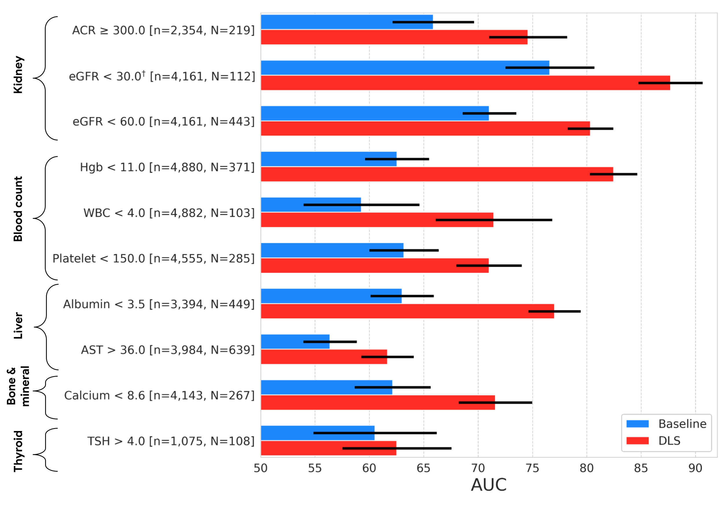
, we compared our external eye design to a standard design (a logistic regression design taking clinicodemographic variables as input) by calculating the location under the receiver operator curve (AUC). The AUC varies from 0 to 100%, with 50% suggesting random efficiency and greater worths suggesting much better efficiency. For all however among the 9 forecast jobs, our design statistically surpassed the standard design. In regards to outright efficiency, the design’s AUCs varied from 62% to 88%. While these levels of precision are most likely inadequate for diagnostic applications, it remains in line with other preliminary screening tools, like mammography and pre-screening for diabetes, utilized to assist determine people who might gain from extra screening. And as a non-invasive available method, taking pictures of the external eye might use the possible to assist screen and triage clients for confirmatory blood tests or other medical follow-up.
Outcomes on the EyePACS test set, revealing AUC efficiency of our DLS compared to a standard design. The variable “n” describes the overall variety of datapoints, and “N” describes the variety of positives. Mistake bars reveal 95% self-confidence periods calculated utilizing the DeLong technique
 |
| â Shows that the target was pre-specified as secondary analysis; all others were pre-specified as main analysis. The external eye pictures utilized in both this and the previous research study were gathered utilizing table leading cams that consist of a head rest for client stabilization and produce high quality images with excellent lighting. Considering that image quality might be even worse in other settings, we wished to check out to what level the DLS design is robust to quality modifications, beginning with image resolution. Particularly, we scaled the images in the dataset to a series of sizes, and determined efficiency of the DLS when re-trained to manage the downsampled images. Listed below we reveal a choice of the outcomes of this experiment (see the |
paper
for more total outcomes). These outcomes show that the DLS is relatively robust and, for the most part, outshines the standard design even if the images are reduced to 150×150 pixels. This pixel count is under 0.1 megapixels, much smaller sized than the common mobile phone electronic camera. Impact of input image resolution. Leading:
 |
| Sample images scaled to various sizes for this experiment. Bottom: Contrast of the efficiency of the DLS ( red) experienced and assessed on various image sizes and the standard design ( blue). Shaded areas reveal 95% self-confidence periods calculated utilizing the DeLong technique. Conclusion and future instructions Our previous research study showed the guarantee of the external eye method. In this work, we carried out a more extensive search to determine the possible systemic biomarkers that can be anticipated from these pictures. Though these outcomes are appealing, lots of actions stay to figure out whether innovation like this can assist clients in the real life. In specific, as we point out above, the images in our research studies were gathered utilizing big tabletop cams in a setting that managed aspects such as lighting and head positioning. Moreover, the datasets utilized in this work consist mostly of clients with diabetes and did not have adequate representation of a variety of essential subgroups– more concentrated information collection for DLS improvement and examination on a more basic population and throughout subgroups will be required prior to thinking about medical usage. |
We are thrilled to check out how these designs generalize to mobile phone images provided the possible reach and scale that this allows for the innovation. To this end, we are continuing to deal with our co-authors at partner organizations like
Chang Gung Memorial Medical Facility
in Taiwan, Aravind Eye Medical Facility in India, and EyePACS in the United States to gather datasets of images recorded on smart devices. Our early outcomes are appealing and we eagerly anticipate sharing more in the future.
Recognitions This work included the efforts of a multidisciplinary group of software application engineers, scientists, clinicians and cross practical factors. Secret factors to this task consist of: Boris Babenko, Ilana Traynis, Christina Chen, Preeti Singh, Akib Uddin, Jorge Cuadros, Lauren P. Daskivich, April Y. Maa, Ramasamy Kim, Eugene Yu-Chuan Kang, Yossi Matias, Greg S. Corrado, Lily Peng, Dale R. Webster, Christopher Semturs, Jonathan Krause, Avinash V Varadarajan, Naama Hammel and Yun Liu. We likewise thank Dave Steiner, Yuan Liu, and Michael Howell for their feedback on the manuscript; Amit Talreja for examining code for the paper; Elvia Figueroa and the Los Angeles County Department of Health Solutions Teleretinal Diabetic Retinopathy Evaluating program personnel for information collection and program assistance; Andrea Limon and Nikhil Kookkiri for EyePACS information collection and assistance; Dr. Charles Demosthenes for drawing out the information and Peter Kuzmak for getting images for the VA information. Finally, an unique thanks to Tom Small for the animation utilized in this post.